The inner eye includes the iris, the anterior chamber and the lens. The most common condition that owners notice themselves is cataract – a clouding of the lens that can lead to blindness.
When you should see us
- a greyish or whitish cloudiness in the pupil
- a sudden deterioration in vision, for example bumping into furniture or uncertainty in the dark
- your pet has diabetes – diabetes can lead to cataract very quickly
- a red, painful eye with a small pupil as a possible sign of inflammation inside the eye (uveitis)
What we examine
We assess the lens, iris and anterior chamber with the slit lamp and check for inflammation inside the eye or a displaced lens. Before any possible surgery, we also use ultrasound and a retinal function test (electroretinogram) to check whether the retina behind the clouded lens is still intact – this is decisive for whether surgery can really benefit vision.
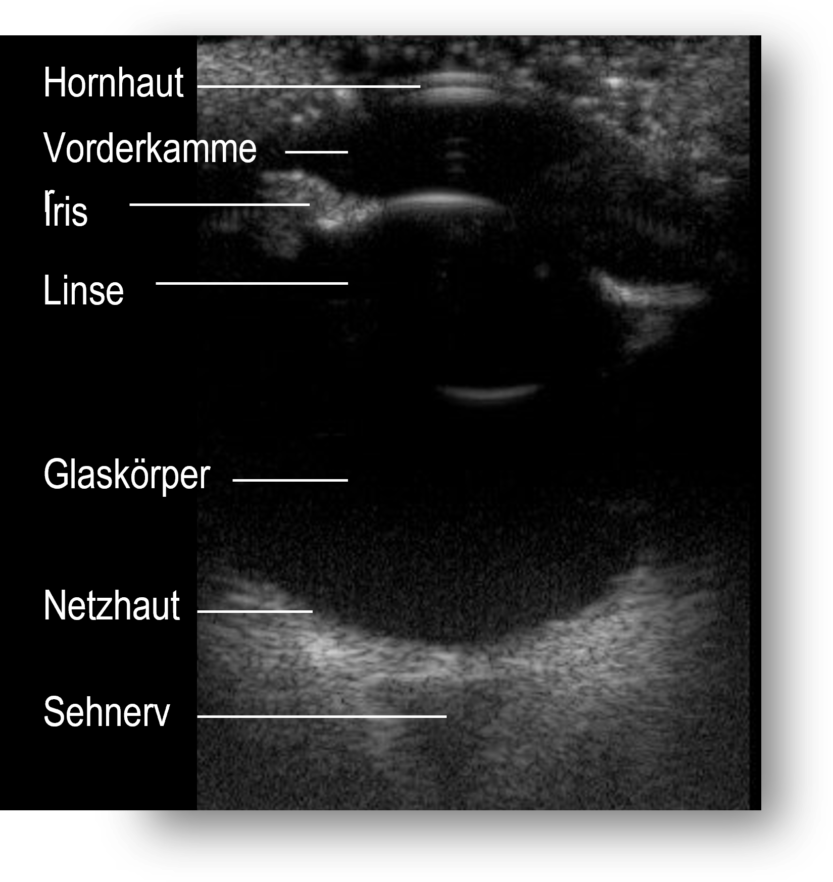
Ocular ultrasound: if the inside of the eye can no longer be seen directly because of the clouding, we use ultrasound to check whether the retina and vitreous are normal.
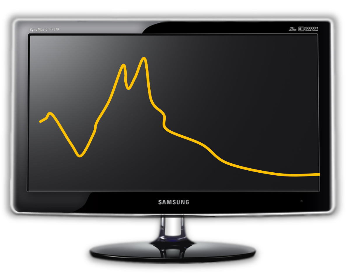
Electroretinogram (ERG): this retinal function test shows whether surgery can benefit vision at all.
Cataract surgery
We perform cataract surgery – removing the clouded lens and implanting an artificial lens – at our practice in Greifswald. Whether and when surgery makes sense for your pet, we discuss with you in detail after the preliminary examination: the stage of the cataract, general health and the result of the retinal function test are the most important factors.
More about the different forms and causes of cataract can be found in our knowledge base (in German).