The cornea is the clear, sensitive front surface of the eye. If it is injured, inflamed or becomes cloudy, this usually shows as a red, watering or squinted eye.
When you should see us
- a suddenly painful, watering or squinted eye
- visible cloudiness or a whitish, greyish or dark spot on the cornea
- a corneal defect that does not heal despite treatment by your family vet
- chronic, sticky eye discharge or recurring conjunctivitis
Common conditions
- injuries and ulcers of the cornea, including deep or already perforated defects
- persistent, non-healing defects such as SCCED (“boxer ulcer”)
- chronic inflammation, for example the hereditary pannus disease in German Shepherds
- infections, among others with feline herpesvirus
- cornea nigra (corneal sequestrum) – a blackish tissue necrosis that occurs mainly in short-nosed cat breeds
- dermoids – congenital, hairy islands of skin on the cornea that rub constantly
- dry eye and foreign bodies
What we examine
We stain the corneal surface with a special dye (fluorescein) – defects show up green, even when they are barely visible to the naked eye.
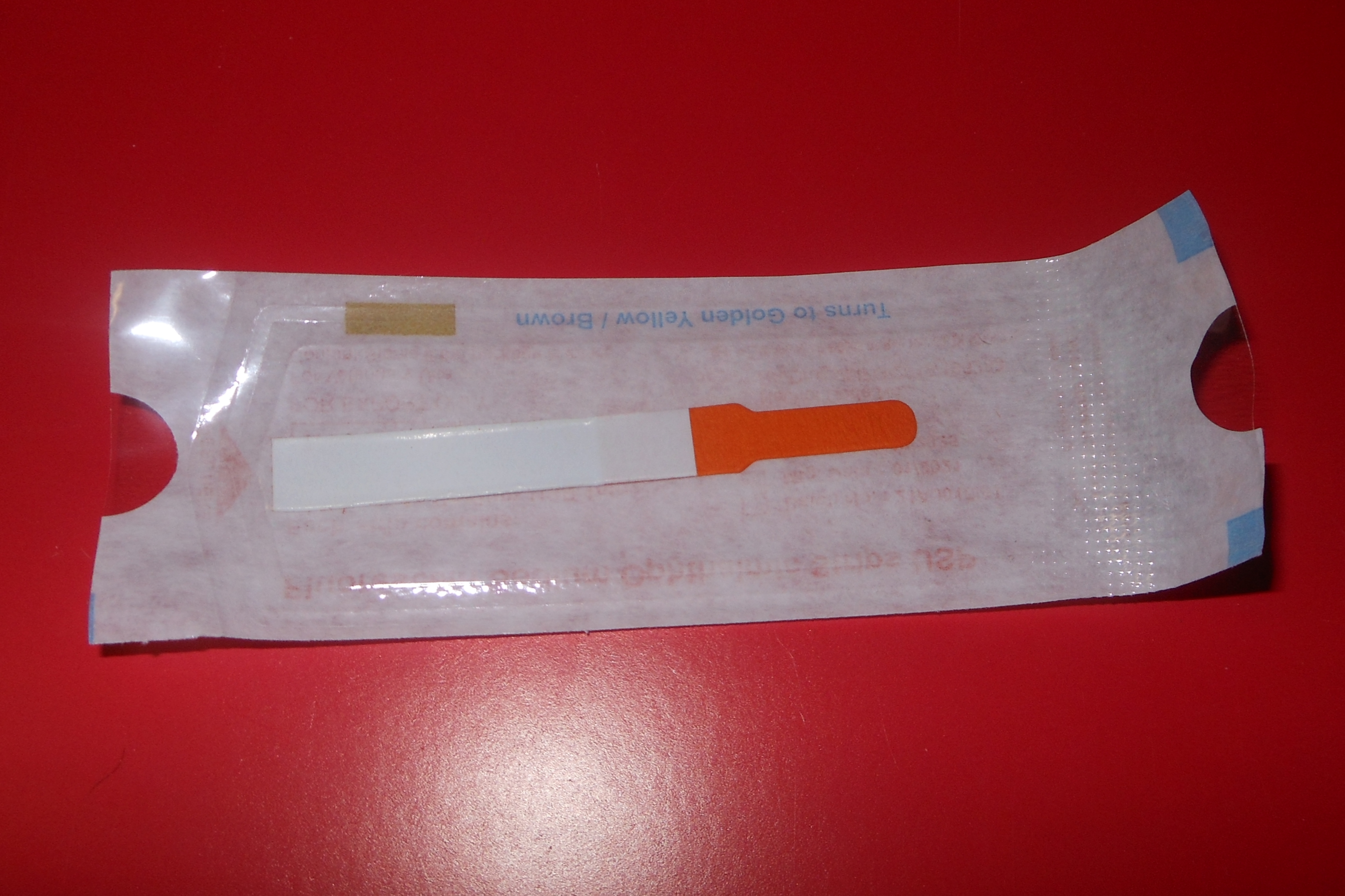
Fluorescein test strips for staining the corneal surface.
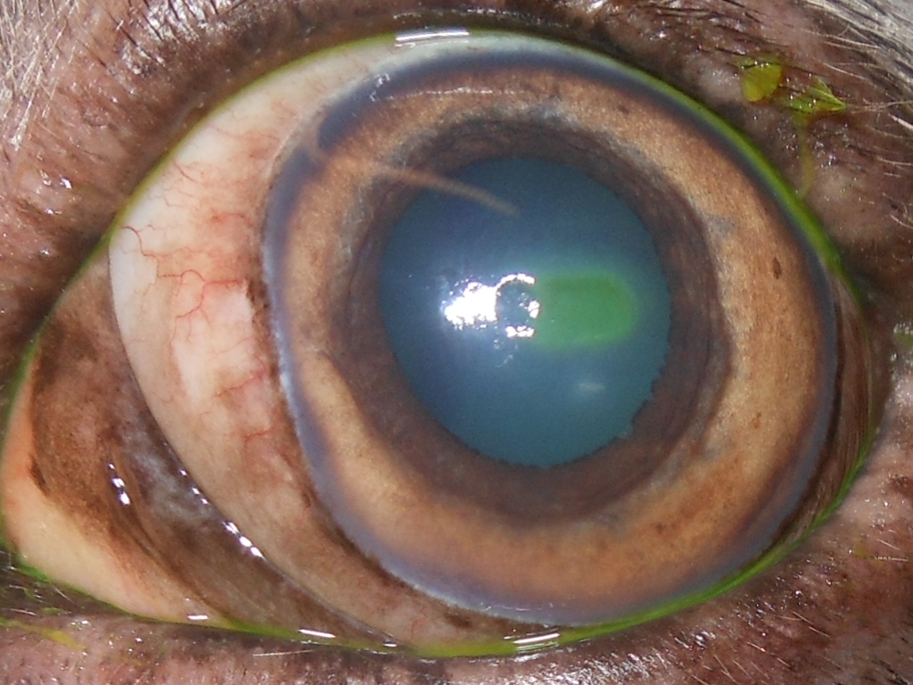
Corneal defect stained with fluorescein – the dye makes the wound clearly visible.
Treatment
Depending on the findings, treatment ranges from eye drops and a minor procedure under local anaesthesia – for example roughening the wound edges of non-healing defects, sometimes with a fine diamond burr (corneal debridement) – to surgery under general anaesthesia. For deep or already perforated defects we cover the cornea with the animal’s own tissue (conjunctival or corneoconjunctival graft) or stabilise it temporarily with a special contact lens or an amniotic membrane product until the wound can bear the load again on its own.
More about individual corneal conditions – such as the melting corneal ulcer or herpes infection in cats – can be found in our knowledge base (in German).